
Protecting our bones after diabetes and hypertension
Researchers used the CLS to identify a potential bone health therapy.
By Colleen MacPherson.Bone X-ray.
On November 14, World Diabetes Day aims to raise awareness for the global health threat posed by diabetes, which affects over 460 million people globally, and to promote coordinated efforts to confront diabetes.
People living with type II diabetes and hypertension face an increased risk of bone fractures. An international team of researchers has used the Canadian Light Source (CLS) at the University of Saskatchewan (USask) to identify a potential bone health therapy that could one day alleviate that problem.
The collaboration between the Bone-Muscle Research Center at the University of Texas at Arlington (BMRC-UTA) and the Colleges of Medicine and Kinesiology at USask explored whether hepatocyte growth factor (HGF) could help reduce the fracture risk for people with type II diabetes. Since 50-85 % of diabetic patients live with hypertension, and both conditions are linked to a higher risk of breaks, this population is particularly vulnerable.
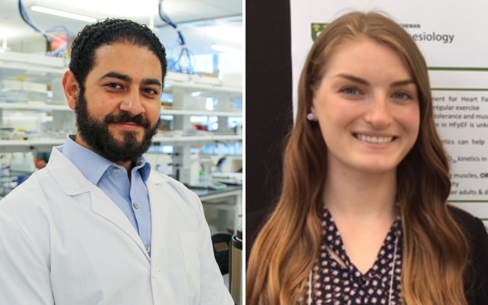
Dr. Kamal Awad, research scientist at the BMRC-UTA and first author on the study, said “bones protect our internal organs and allow us to move, thus maintaining a healthy bone is crucial especially for people suffering from diabetes and hypertension”.
This study focused on HGF, which is a naturally occurring molecule that is known to regulate cell growth throughout the body. Awad said it is also “associated with bone regeneration, remodelling, and the balance between osteoblast and osteoclast, but what was unknown is how HGF affects the chemical structure of the bone.”
Natasha Boyes, a PhD candidate specializing in cardiovascular disease in the College of Kinesiology at USask and first co-author, is interested in the whole-body effects of cardiovascular disease, and explained remodelling as a change process bones undergo throughout a person’s life.
“Most people think bone should be hard,” she said, “but hard bone can be very brittle. What you want is bone with the right architecture, and bone is always changing. Any stimulus can cause bone to adjust its structure. For example, if you’re a runner, your bones will change and adapt to better cope with the pounding (biomechanical stress). That’s remodelling.”
To explore how HGF might improve bone health, the researchers did site-specific injections of HGF on diabetic hypertensive rats, then used spectroscopy at the CLS to study the bone chemical structure with a focus on calcium and phosphorous. The team utilized the facility’s specialized SGM, VLS-PGM, and SXRMB beamline facilities for this analysis.
The results of the study, published in the Journal of Materials Research, showed increases in soluble phosphate in the treated bones. This change indicates that HGF may stimulate the bone into a remodelling phase in response to the detrimental effects of diabetes, hypertension, and the drugs used in treatment.
Awad said synchrotron techniques are critical to understanding the exact chemical structure or the coordination of the calcium and phosphate in bones, but further research is required. Dr. Venu Varanasi, lead corresponding author, indicated that his team is expanding the collaboration with the USask team lead by Dr. Corey Tomczak to investigate what exactly what HGF does in bone remodelling and how to fine tune its effects as a potential therapy.
For both Boyes and Awad, the long-term goal is to develop a therapy to reduce bone fracture risk, especially in people with type II diabetes and hypertension. Awad added future HGF therapies may also have a role to play in speeding up the healing process after procedures like bone grafts.
Awad, Kamal, Natasha G. Boyes, Ramlah Iqbal, Mohamed Ahmed, Adel Mohamed, Pranesh Aswath, Corey R. Tomczak, and Venu Varanasi. "Hepatocyte growth factor administration increases bone soluble phosphate and alters bone chemical structure in diabetic hypertensive rats." Journal of Materials Research (2021): 1-16.
Photos: Kamal Awad | Natasha Boyes | CLS synchrotron
Media Relations:
Victoria Martinez
Communications Coordinator
Canadian Light Source
306-657-3771
Victoria.martinez@lightsource.ca