
X-ray Diffraction
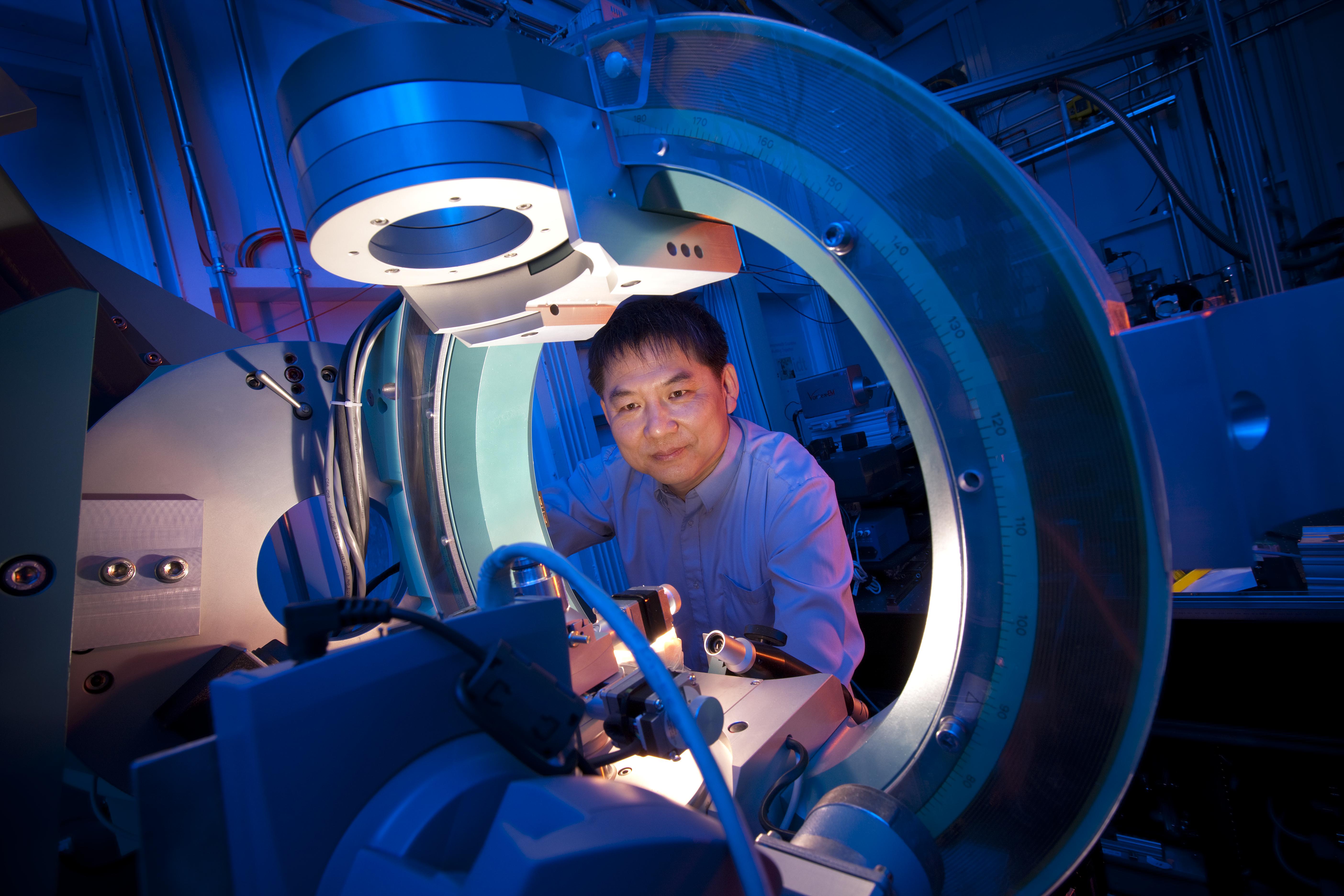
X-ray diffraction (XRD) is a technique that can be used to study the structure of materials. The atoms of a material form a crystalline structure and, by virtue of their uniform spacing, cause an interference pattern in an incident beam of X-rays. Variations of XRD can be used to study different sample types and conditions, including Liquid XRD, Grazing Incidence XRD, Reciprocal Space Mapping, or Anomalous XRD.
Energy Range: 2.7-94 keV
Soft X-ray Energy Range: 80-2,000 eV
Applications:
CATALYSTS DRUG DEVELOPMENT
Loading...
MX
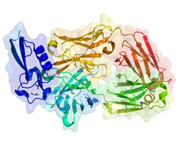
Macromolecular Crystallography (MX) is the most used technique by private-sector clients at the CLS. MX provides high-resolution structural models of proteins and other large biomolecules, allowing for targeted drug design and development. Our crystallography facilities are highly automated, producing excellent-quality data with fast throughput (up to 12 datasets per hour). Samples are typically shipped to the CLS where data can be collected remotely (or you can have one of our experts collect it for you).
Energy Range: 4-22 keV
Applications:
CROP DEVELOPMENT DRUG DEVELOPMENT
Loading...
PXRD
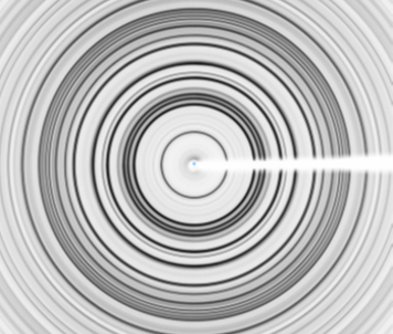
Powder X-ray Diffraction is a workhorse technique in many industries, providing direct quantitative information about crystal structures, phase composition, stress/strain, and crystallite size. Synchrotron PXRD provides significantly greater sensitivity and spectral resolution compared to conventional instruments, allowing for quantification of very dilute phases and determination of fine structural details. PXRD is a rapid-access analytical service, and in many cases we're able to turn around sets of samples within a couple of weeks.
Energy Range: 4-94 keV
Applications:
CATALYSTS DRUG DEVELOPMENT ENERGY STORAGE OIL & GAS MINING
Loading...
SAXS
Small Angle X-ray Scattering (SAXS) experiments provide nanoparticle size distributions, information on the size and shape of macromolecules, and determine characteristic distances of partially ordered materials by analyzing the scattering behaviour of x-rays passing through a sample. These x-rays are measured at very small angles, typically between 0.1 and 10°.
Applications:
CATALYSTS DRUG DEVELOPMENT
Loading...
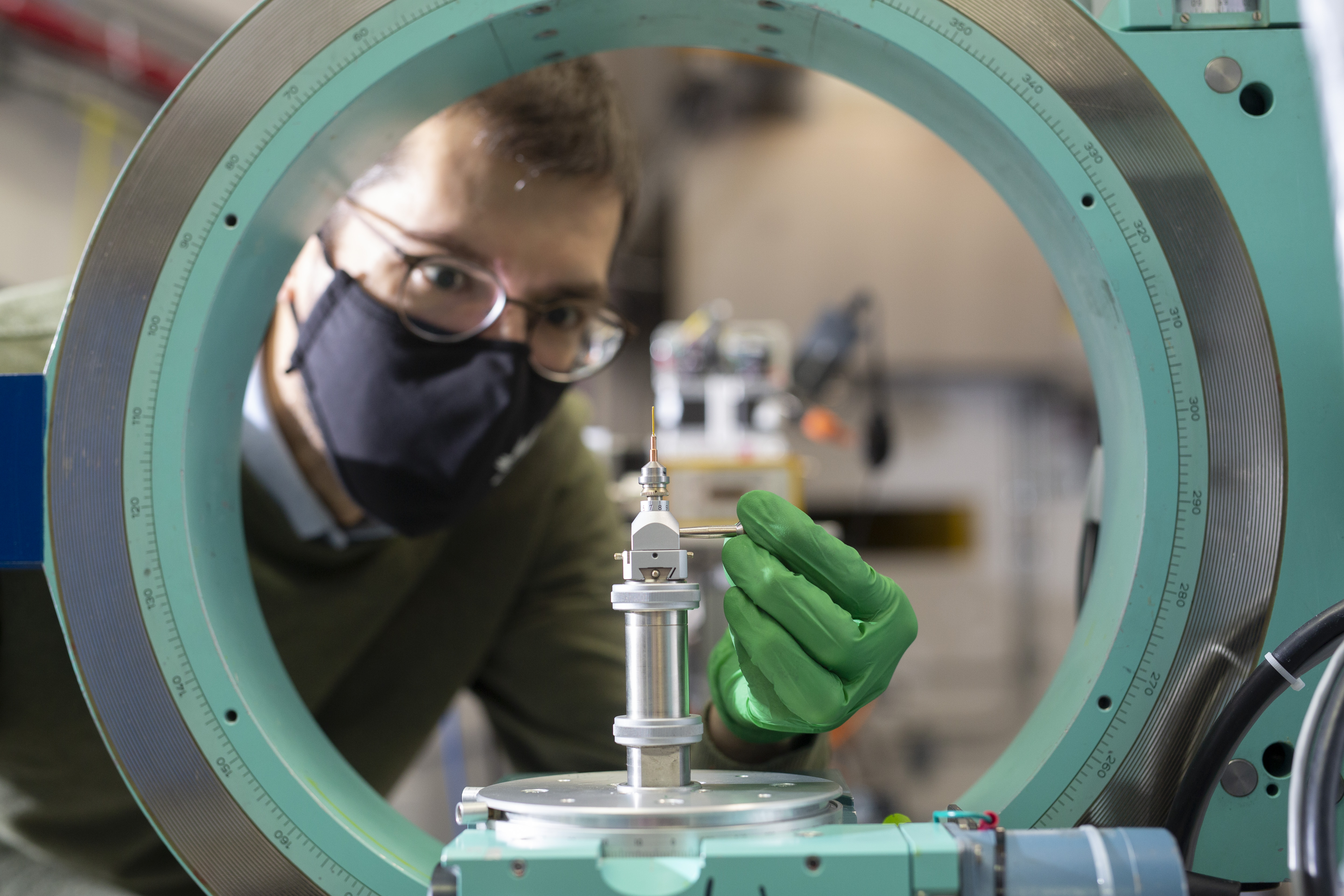
Small Molecule X-ray Diffraction
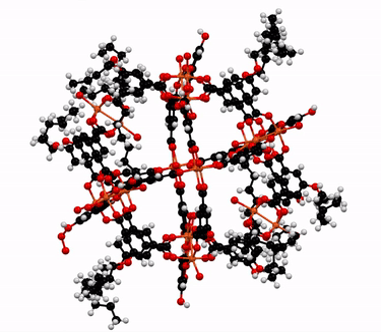
X-ray crystallography has long been the fundamental technique used by chemists to determine 3D structures of organic and inorganic compounds. Synchrotrons provide high-resolution structural models without the requirement of large single crystals and long acquisition times that many laboratory instruments require. This technique is very complimentary to other structural techniques and can provide important information on the arrangement and orientation of organic structures and functional groups.
Applications:
CATALYSTS DRUG DEVELOPMENT
Loading...
Resonant X-ray Scattering
Resonant x-ray scattering can be measured with a spectrometer during X-ray Emission Spectroscopy (XES) experiments. As electrons are excited out of their shells, the emitted photons of the recombinating electrons are measured. Inelastic scattering appears relative to an elastic emission line. This scattering is both element- and site-specific, making it a powerful tool for determining detailed electronic properties of materials.
Energy Range: 2.7-94 keV
Soft X-ray Energy Range: 80-2,000 eV
Applications:
CATALYSTS DRUG DEVELOPMENT
Loading...
Access
Purchased Access
Purchased access offers quick and accurate solutions to proprietary questions. CLS scientists develop experimental plans based on clients' needs, collect and analyze data, and provide detailed reports with key answers to critical questions.
Peer-Reviewed Access
Academic clients can submit proposals through a peer review process. Beam time is granted based on scientific merit, with the expectation that any results will be published. In special cases, rapid access is also available for instrument or beam time.