
Computed Tomography
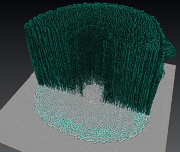
Computed Tomography (CT) is a common x-ray imaging technique (especially in medicine), where a full 3D image is acquired that shows the complete internal structure of a sample. Synchrotron CT provides much higher contrast than clinical CT, enough to resolve very similar materials (like a carbon fiber in a resin matrix). Synchrotron CT is also many orders of magnitude faster than conventional: a full scan at a resolution of several microns can be completed in a matter of seconds as opposed to hours.
Energy Range: 6-150 keV
Applications:
CROP DEVELOPMENT ENERGY MATERIALS CATALYSTS BIOMEDICAL IMAGING OIL & GAS
Loading...
FTIR Spectromicroscopy
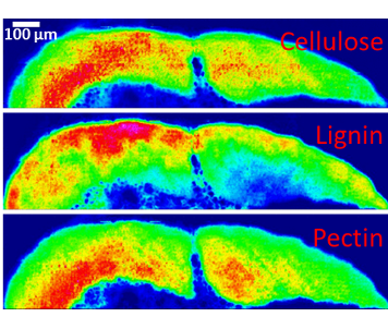
IR-based spectroscopic techniques (like FTIR and Raman spectroscopy) are common laboratory techniques that provide useful information about the composition of organic samples (including identification of functional groups). Bench-top IR microscopes exist as well, allowing for spectromicroscopic mapping of IR-active samples, but synchrotron-based IR maps provide better signal-to-noise and higher resolution (down to the diffraction limit near 1 micron) compared to lab-based systems.
Energy Range: .0062 - 0.744 cm-1
Applications:
CROP DEVELOPMENT CROP DISEASE ENERGY MATERIALS BIOMEDICAL IMAGING
Loading...
X-ray Spectromicroscopy
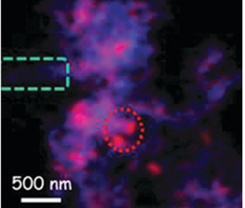
X-ray spectroscopic imaging (also known as spectromicroscopy or SM) involves collecting an X-rays absorption spectrum using a beam that is focused down to a very small spot. This spot is scanned over a sample, generating an image where each pixel contains a complete chemical spectrum for a particular element. The spectrum can be a fluorescence spectrum (which shows the distribution of elements) or an X-ray absorption spectrum (which gives chemical information like oxidation states or functional groups). This powerful technique can be performed at resolutions of a couple of millimeters (pixel dimensions) down to as low as 20 nanometers.
Energy Range: 130-2700eV
Applications:
AGRI-PRODUCTS CATALYSTS BIOMEDICAL IMAGING
Loading...
XRF Imaging
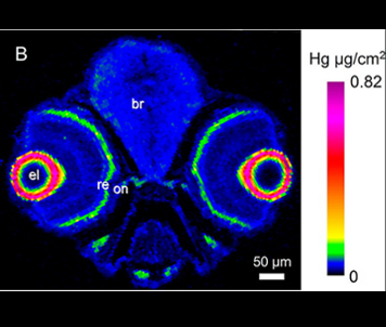
X-ray fluorescence (XRF) is a common laboratory technique for determining the relative amounts of different elements present in a sample. By focusing a synchrotron beam down to a very small spot and measuring the resulting XRF pattern given off by the sample, an elemental map can be created for multiple elements simultaneously. These maps are highly sensitive and are extremely useful for correlating the distribution of elements in a sample.
Energy Range: 4.3-50 keV
Applications:
AGRI-PRODUCTS CATALYSTS BIOMEDICAL IMAGING
Loading...
Access
Purchased Access
Purchased access offers quick and accurate solutions to proprietary questions. CLS scientists develop experimental plans based on clients' needs, collect and analyze data, and provide detailed reports with key answers to critical questions.
Peer-Reviewed Access
Academic clients can submit proposals through a peer review process. Beam time is granted based on scientific merit, with the expectation that any results will be published. In special cases, rapid access is also available for instrument or beam time.