Drug Development
Drug development and protein crystallography are one of the core applications of synchrotron facilities.
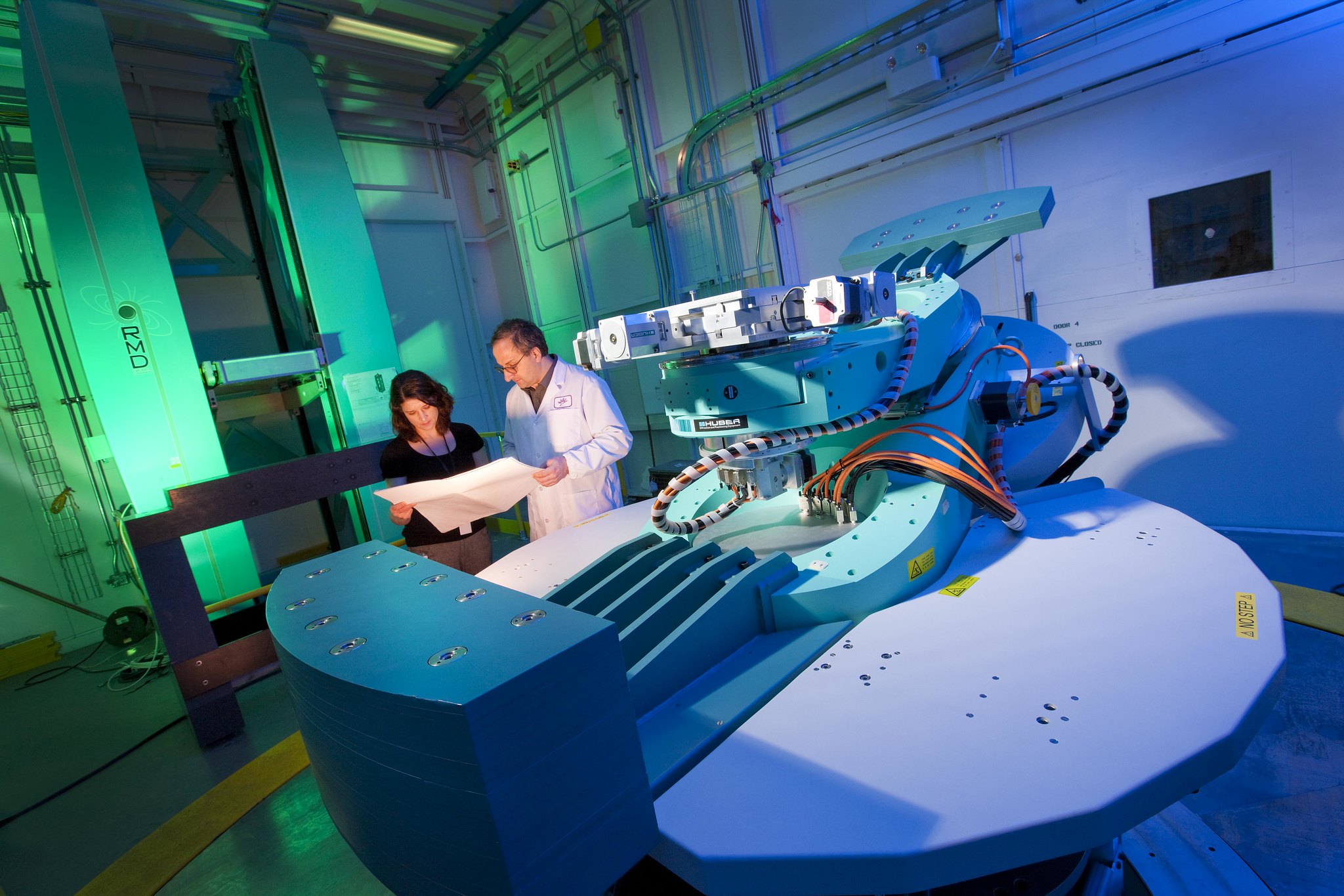
The primary technique used by our pharmaceutical clients is macromolecular crystallography (MX). MX provides detailed protein structures at angstrom-level spatial precision. Knowing the exact structures of target proteins and enzymes allows for the rational design of drugs and cofactors that interact with the active sites of target macromolecules. Aside from MX, we also offer analytical services for small molecule and powder X-ray diffraction (PXRD), which can provide detailed structures of organic molecules, including pharmaceuticals. Synchrotron techniques can provide an important technical basis for creating pharmaceutical intellectual property (IP), and can be critical for determining the outcome of patent litigation. We have provided patent litigation and IP consulting services, and have a great deal of experience in drug development, so contact us today to see how we can help!
Techniques:
MACROMOLECULAR CRYSTALLOGRAPHY (MX) POWDER X-RAY DIFFRACTION (PXRD) SMALL ANGLE X-RAY SCATTERING (SAXS) SMALL MOLECULE X-RAY DIFFRACTIONKetcham, J. M., Harwood, S. J., Aranda, R., Aloiau, A. N., Bobek, B. M., Briere, D. M., Burns, A. C., Caddell Haatveit, K., Calinisan, A., Clarine, J., Elliott, A., Engstrom, L. D., Gunn, R. J., Ivetac, A., Jones, B., Kuehler, J., Lawson, J. D., Nguyen, N., Parker, C., … Marx, M. A. (2024). Discovery of pyridopyrimidinones that selectively inhibit the H1047R pi3kα mutant protein. Journal of Medicinal Chemistry, 67(6), 4936–4949. https://doi.org/10.1021/acs.jmedchem.4c00078
Chew, K. S., Wells, R. C., Moshkforoush, A., Chan, D., Lechtenberg, K. J., Tran, H. L., Chow, J., Kim, D. J., Robles-Colmenares, Y., Srivastava, D. B., Tong, R. K., Tong, M., Xa, K., Yang, A., Zhou, Y., Akkapeddi, P., Annamalai, L., Bajc, K., Blanchette, M., … Kariolis, M. S. (2023). CD98HC is a target for brain delivery of Biotherapeutics. Nature Communications, 14(1). https://doi.org/10.1038/s41467-023-40681-4
Wilding, B., Scharn, D., Böse, D., Baum, A., Santoro, V., Chetta, P., Schnitzer, R., Botesteanu, D. A., Reiser, C., Kornigg, S., Knesl, P., Hörmann, A., Köferle, A., Corcokovic, M., Lieb, S., Scholz, G., Bruchhaus, J., Spina, M., Balla, J., … Neumüller, R. A. (2022). Discovery of potent and selective HER2 inhibitors with efficacy against HER2 exon 20 insertion-driven tumors, which preserve wild-type EGFR signaling. Nature Cancer, 3(7), 821–836. https://doi.org/10.1038/s43018-022-00412-y
Karagiannis, A., Tyryshkin, A. M., Lalancette, R. A., Spasyuk, D. M., Washington, A., & Prokopchuk, D. E. (2022). A redox-active Mn(0) dicarbene metalloradical. Chemical Communications, 58(93), 12963–12966. https://doi.org/10.1039/d2cc04677f
Pushie, M. J., Summers, K. L., Nienaber, K. H., Pickering, I. J., & George, G. N. (2022). Synthesis and structural characterization of copper–cuprizone complexes. Dalton Transactions, 51(27), 10361–10376. https://doi.org/10.1039/d2dt01475k
Chen, Shuo; Wu, Jia-Le; Liang, Ying; Tang, Yi-Gang; Song, Hua-Xin; Wu, Li-Li; Xing, Yang-Fei; Yan, Ni; Li, Yun-Tong; Wang, Zheng-Yuan; Xiao, Shu-Jun; Lu, Xin; Chen, Sai-Juan and Lu, Min. (2021). Arsenic trioxide rescues structural p53 mutations through a cryptic allosteric site. Cancer Cell 39(2), 225-239. 10.1016/j.ccell.2020.11.013
Loading...
Try our MX program!
Biomedical Imaging

X-ray imaging is a staple of clinical and biomedical R&D. Synchrotron radiography and CT take X-ray imaging to a whole new level.
X-ray imaging is one of the most ubiquitous medical imaging tools available. Clinical X-ray imaging can only be used for high-contrast systems such as bone/tissue or heavy-element contrast agents, and the images are typically low-resolution (with pixels on the scale of mm). Synchrotron X-ray imaging offers significantly higher contrast, allowing for the imaging of soft tissue like organs, vasculature, and tumors - often without the need for a contrast agent. Synchrotron CT is also considerably faster, allowing for micron-level imaging at high speed (often whole CT scans can be collected in a matter of seconds). This is especially useful for in-vivo and in-situ applications. Industrial applications include medical implants, research animals, excised tissues, dental structures and much more!
Techniques:
COMPUTED TOMOGRAPHYHedayat, Assem; Belev, George; Zhu, Ning; Bond, Toby and Cooper, David. (2018). Investigating the presence of mercury under a dental restoration using synchrotron K-edge subtraction imaging. Microscopy and Microanalysis, 24(2), 362-363. 10.1017/S1431927618014101
Loading...
Case Studies
Loading...
