
Promising new approach to rebuild bone tissue
McGill University researchers used the CLS to advance a novel method for growing synthetic bone tissue.
By Greg BaskyOver the last 30 years, the scientific community has been working to develop a synthetic alternative to bone grafts for repairing diseased or damaged bone. McGill University researchers used the Canadian Light Source (CLS) at the University of Saskatchewan to advance a novel method for growing synthetic bone tissue.
The rapidly advancing field of bone tissue engineering is focused on growing bone cells in the lab on materials called scaffolds, then transferring these structures into a person’s body to repair bone damage. Like the bone it mimics, scaffolds need an interconnected network of small and large pores that allow cells and nutrients to spread and help generate new bone tissue.
The McGill team’s promising process works by modifying the internal structure of a material, called graphene oxide, to make it more conducive to regenerating bone tissue.
Graphene oxide is an ultrathin, extra strong compound that is being used increasingly in electronics, optics, chemistry, energy storage, and biology. One of its unique properties is that when stem cells are placed on it, they tend to transform into bone-generating cells called osteoblasts.
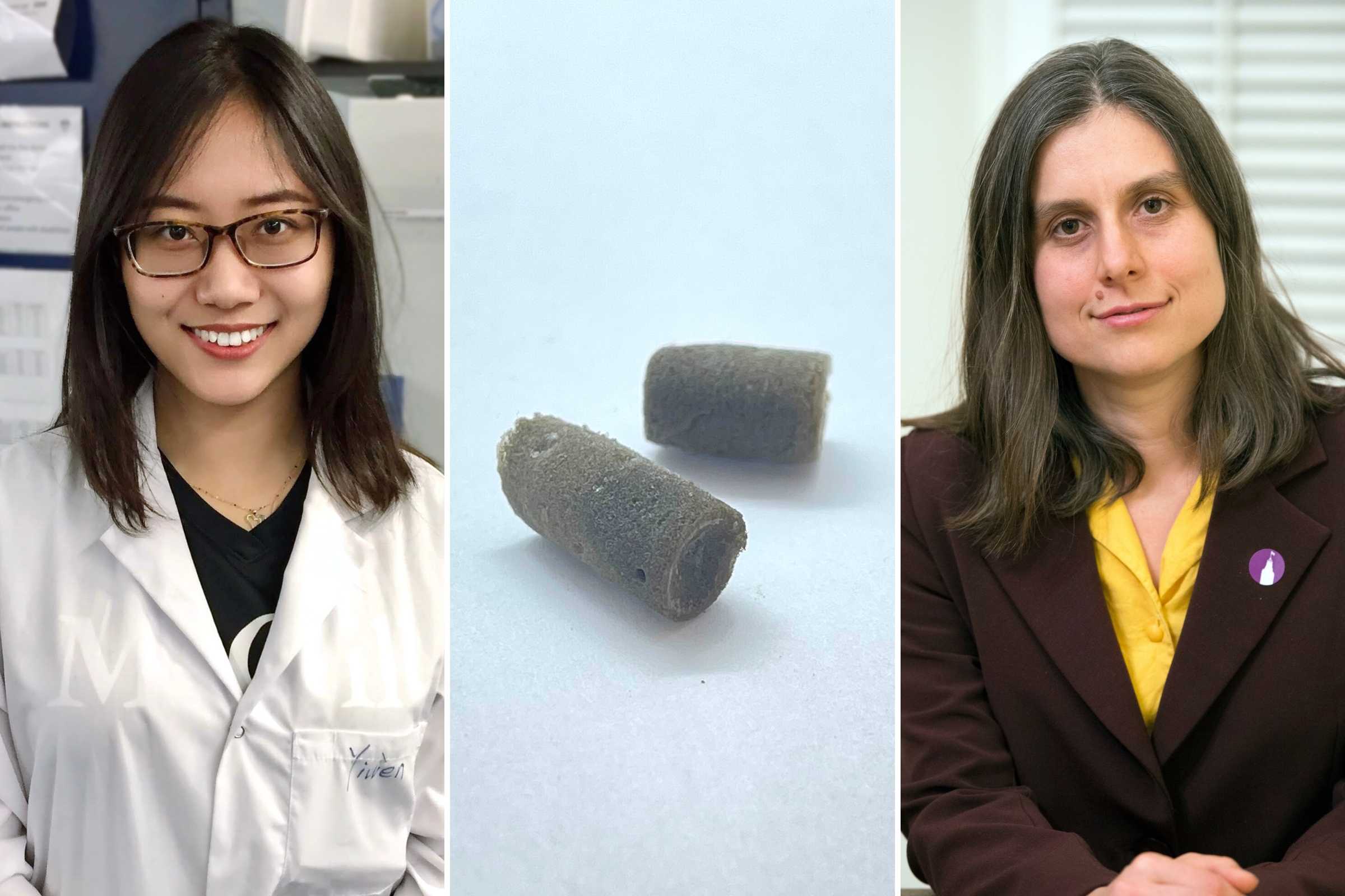
The multidisciplinary group – comprising researchers from McGill’s Departments of Mining and Materials Engineering, Electrical Engineering, and Dentistry – found that adding an emulsion of oil and water to the graphene oxide, then freezing it at two different temperatures, yielded two different sizes of pores throughout the material.
Professor Marta Cerruti said that when they “seeded” the now-porous scaffolding with stem cells from mouse bone marrow, the cells multiplied and spread inside the network of pores, a promising sign the new approach could eventually be used to regenerate bone tissue in humans.
“We showed that the scaffolds are completely biocompatible, that the cells are happy when you put them in there, and that they’re able to penetrate all through the scaffold and colonize the whole scaffold,” she stated.
The researchers used the BMIT-BM beamline at the CLS to visualize the different sized pores inside the scaffolding as well as the growth and spread of the cells. Lead researcher Yiwen Chen, a PhD student working under Cerruti, said their work would not have been possible without the synchrotron because the low density of graphene oxide means it absorbs only a very small amount of light.
“To our knowledge, this is the first time that people have used synchrotron light to see the structure of graphene oxide scaffolds,” said Chen.
While widespread clinical application of this new approach may still be many years away, Cerruti thinks their work could enable other researchers to learn more about how stem cells morph into bone cells.
“Maybe this will lead to a better understanding of the biology of bones that we wouldn't understand otherwise,” she said. “Perhaps in the shorter term we can use the methods in the lab to better understand bone and perhaps develop new drugs.”
Chen, Yiwen, Xinyun Su, Dominic Esmail, Emily Buck, Simon D. Tran, Thomas Szkopek, Marta Cerruti. “Dual-templating strategy for the fabrication of graphene oxide, reduced graphene oxide and composite scaffolds with hierarchical architectures.” Carbon 189 (2022): 186-198 https://doi.org/10.1016/j.carbon.2021.12.054
Photos: Synchrotron | BMIT beamline | Cerruti, Chen, and scaffolds
Media Relations:
Victoria Schramm
Communications Coordinator
Canadian Light Source
306-657-3516
victoria.schramm@lightsource.ca