
BMIT - Biomedical Imaging and Therapy Facility
The BMIT facility consists of two beamlines (low-energy BMIT-BM and high-energy BMIT-ID beamline). It is dedicated to imaging of biological tissues and radiation therapy research. New instruments are successfully applied for general-purpose X-ray microtomography and imaging of fast processes.
Spectral Range
| BMIT-ID | BMIT-BM |
| 28 - 140 keV | 12.6 – 40.0 keV |
Techniques
Imaging
| BMIT-ID | BMIT-BM |
X-ray Irradiation Experiments/Dosimetry
|
X-ray Irradiation Experiments/Dosimetry
|
Samples
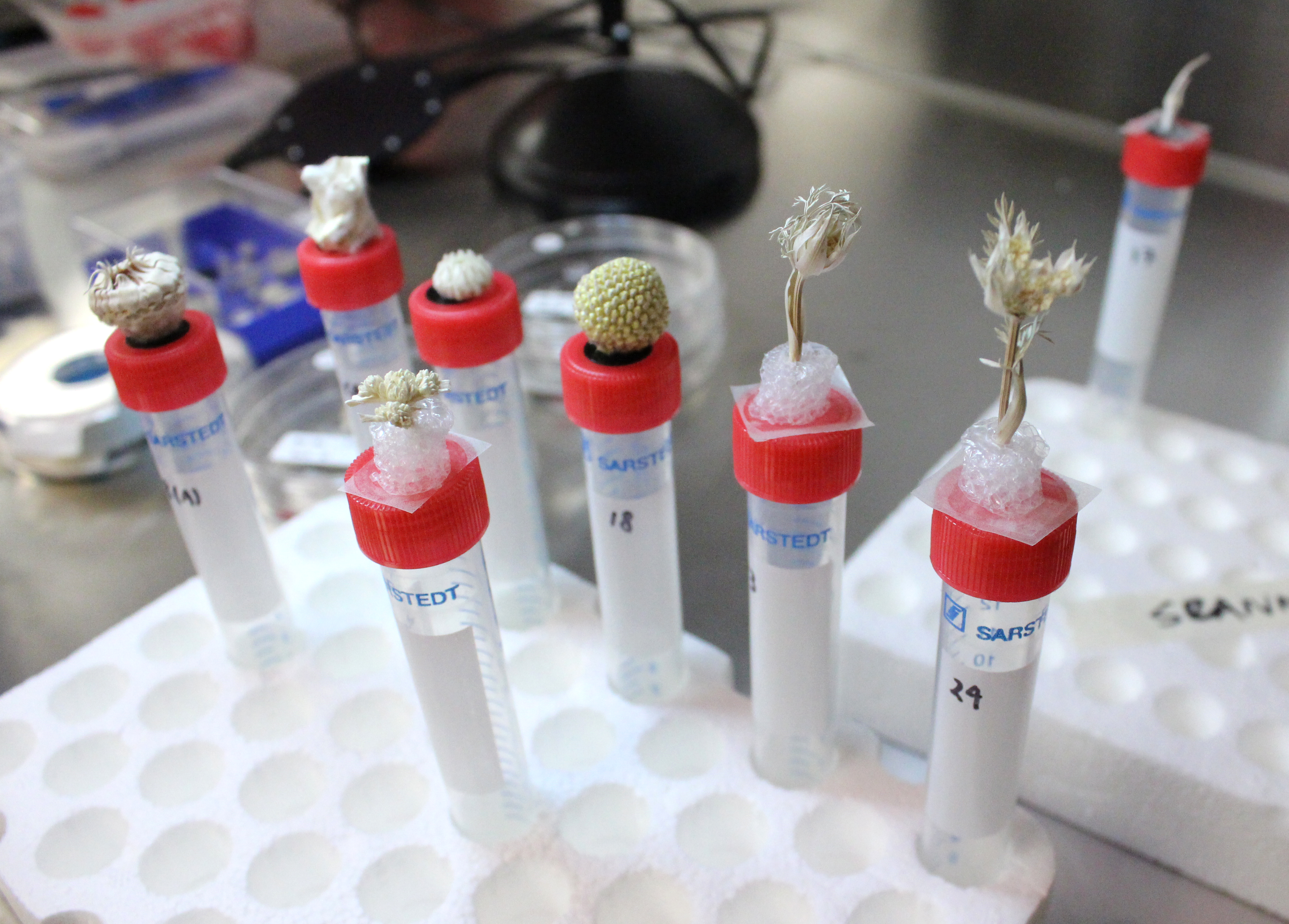
BMIT supports a variety of samples ranging from mm-size tissue and plant specimens to model organisms such as rabbits and small piglets, 20-cm-large fossils, and bulky in-situ rigs.
Ethics approval is required before using some samples, including live animals.
Specifications
|
|
BMIT-ID | BMIT-BM |
| CLS Port | 05ID-2 | 05B1-1 |
| Source | Superconducting Wiggler | Bending Magnet |
| Typical spot sizes | 160 mm x 10 mm @FWHM @ 30 keV, gradually decreases down to 130 m x 6 mm @ 100 keV | BMIT-BM: Horizontal profile is flat with max width of 200 mm; vertical profile is Gaussian-like with 4 mm FWHM for mono beam @ 20 keV and ~3 mm for filtered white beam |
| Maximum Photon Flux | ~5x10¹⁰ ph/s/mm2 | 109 ph/s/mm2 @ 20 keV mono, 1012 ph/s/mm2 peak @ 20 keV filtered white beam |
Contacts
Ning Zhu
Interim Beamline Responsible (BMIT)
Visit the BMIT website for a full list of beamline contacts.
Beamline Video
Access
Purchased Access
Purchased access offers quick and accurate solutions to proprietary questions. CLS scientists develop experimental plans based on clients' needs, collect and analyze data, and provide detailed reports with key answers to critical questions.
Peer-Reviewed Access
Academic clients can submit proposals through a peer review process. Beam time is granted based on scientific merit, with the expectation that any results will be published. In special cases, rapid access is also available for instrument or beam time.