Improving heart disease outcomes for all patients
McGill researchers used the CLS to get one step closer to understanding the origins of arterial calcification, a process that contributes to heart disease.
By Erin MatthewsA closeup of a stethoscope.
A better understanding of arterial calcification
McGill researchers are one step closer to understanding the origins of arterial calcification, a process that contributes to heart disease.
Minerals form naturally in our bones and teeth, but when minerals like calcium phosphate attach to the soft tissues of our vascular system, they can turn the once flexible arteries into stiff vessels that restrict blood flow––increasing the chance of heart attacks or strokes.

Understanding how and why minerals form in soft tissue is crucial for the health of at-risk Canadians, those living with diabetes and chronic kidney disease, as well as seniors.
Data collected on the SXRMB beamline at the Canadian Light Source (CLS) at the University of Saskatchewan has helped further the understanding about where these calcium deposits start.
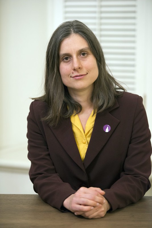
“We have done a lot of work at the CLS on this topic because we have been interested in trying to figure out what minerals are being deposited into the tissues,” lead investigator Marta Cerruti said.
A recent paper published in Crystal Growth and Design is a follow up to previous research conducted at the CLS, which looked at the progression of mineral phases in medial arterial calcification. Cerruti and her collaborators are now focused on understanding exactly where these minerals start to form.
Mineral ions carry a negative or a positive charge and usually deposit on a charged protein. But elastin, the protein in arterial walls, has no overall charge, making it an unusual place for minerals to form.
“In arterial walls you have collagen proteins and elastin proteins––you wouldn’t expect elastin to mineralize,” Cerruti explained, “In bone, calcium phosphate is deposited on collagen. When I first started studying this, I was really surprised that nature would make the minerals form on elastin instead of on collagen.”
To understand this phenomenon, the team focused on a theory first proposed by chemist D.W. Urry in 1974, who suggested that small, uncharged groups on elastin form a kind of pocket that makes mineral deposits on the protein possible.
The McGill team was able to provide new evidence to back up his claim.
Using elastin gels from a natural source, the team’s investigations at the CLS found that minerals were not depositing on the few negatively charged areas of elastin, as other theories have surmised.
“Our results basically told us that, yeah the negatively charged groups are not involved in binding. So, then we looked at the neutral binding sites that Urry was proposing and found that you can correlate neutral groups and mineralization,” Cerruti said.
A better understanding of where mineral deposits form can help in the development of tailored, targeted treatments for arterial calcification in the future, and making heart disease outcomes better for all patients. Currently, there are no treatments for the disease –– mostly due to the limited understanding of its progression.
Zhang, Peng, Ophélie Gourgas, Audrey Laine, Monzur Murshed, Diego Mantovani, and Marta Cerruti. "Coacervation Conditions and Cross-Linking Determines Availability of Carbonyl Groups on Elastin and its Calcification." Crystal Growth & Design 20, no. 11 (2020): 7170-7179. https://doi.org/10.1021/acs.cgd.0c00759
For more information, contact:
Victoria Martinez
Communications Coordinator
306-716-6112
victoria.martinez@lightsource.ca
