Each CLS beamline is unique and provides users with different techniques using synchrotron light. Below you will find information on each of the beamlines and what they can do.
Biological X-ray Absorption Spectroscopy
BioXAS enhances and complements life science research at CLS with three beamlines, two from the wiggler and one from an undulator. Two beamlines are dedicated to XAS (X-Ray Absorption Spectroscopy) and one is a multi-mode XFY imaging line. These systems are tailored for the study of metals in living systems using XAS and XRF imaging. BioXAS investigates the molecular form and microscopic location of metals in biological systems with unprecedented sensitivity.
These studies include investigations of the role of metals in brain diseases like Alzheimer’s, how to treat the deadly effects of toxic elements such as mercury, and developing improved drugs to treat cancer. Environmental research at BioXAS focuses on how metal contaminants affect organisms and, ultimately, humans.
Biomedical Imaging and Therapy
-
Unique in North America, the BMIT facility’s two beamlines will offer advanced imaging for biological tissue in unprecedented detail, as well as high-precision radiation therapies for cancer.
-
Core research programs include human and animal reproduction, cancer imaging and treatment, spinal cord injury and repair, cardiovascular imaging and disease, bone growth and development, arthritis and athletic injuries, dental conditions, mammography, developmental biology, gene expression research, development of new imaging methods as well as extending present imaging capabilities.
BMIT is designed to image biological tissue and to conduct radiation therapy research. The facility will address the interest of scientists and clinicians in the diagnosis and treatment of cancer (breast tumours and paediatric oncology), circulatory and respiratory disease (heart disease and asthma) neurological and behavioural disease (brain and spinal cord injuries, epilepsy), reproductive dysfunction (infertility, menopause, and contraceptives), musculo-skeletal disease and kinesiology (arthritis, athletic injuries), and dental conditions (such as temporomandibular disease). BMIT has two beamlines: one which uses a bending magnet to produce light and one which uses a powerful superconducting wiggler.
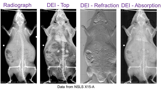
BMIT is one of the few CLS beamlines that produces an X-ray similar to what we generally think of in doctors’ and dentists’ offices. The difference is that BMIT can use different methods to achieve that image. Those differences are important, as can be seen below with an example of Diffraction Enhanced Imaging.
Three experimental endstations at BMIT are capable of using several imaging techniques in both projection and 3D computed tomography (CT) modes. Additionally, the insertion device beamline is capable of microbeam radiation therapy (MRT) and synchrotron stereotactic radiation therapy (SSRT).
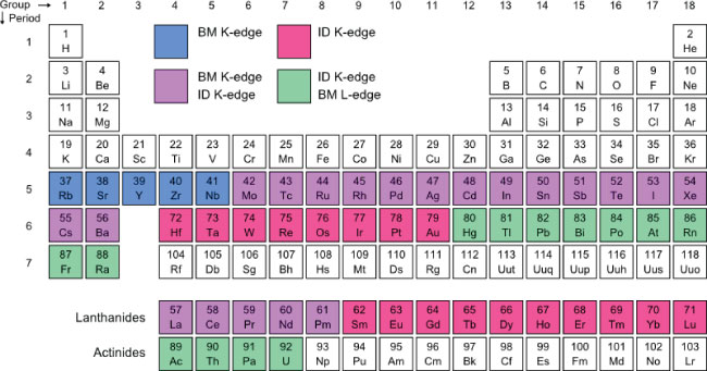
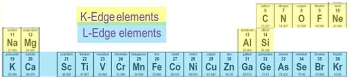
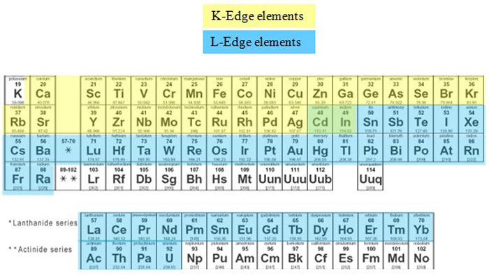
BM -> Bending Magnet
ID -> Insertion Device (Superconducting Wiggler)
Brockhouse X-ray Diffraction and Scattering Sector
- Brockhouse is a suite of 3 beamlines dedicated to using hard X-rays for diffraction and scattering techniques for material science.
The Brockhouse X-Ray Diffraction and Scattering Sector, with three beamlines, is a national centre for structural characterization of many forms of materials systems. This includes crystals, solids, liquids, and nanostructures under ambient conditions and at extreme temperatures, pressures and magnetic fields. The sector will support a diverse community of Canadian and international scientists spanning the disciplines of physics, chemistry, geology, environmental science, biology and engineering. Some potential applications include structural studies of polymers, drugs, emulsions, novel batteries, petroleum products and quantum materials.
Far-Infrared
- This beamline offers wavelengths of light that are very hard to obtain using conventional sources.
- Infrared light causes specific vibrations in molecule bonds, so researchers can identify molecules by their precise vibration pattern.
- Applications include simulations of molecules from space and organic chemistry.
Operating at far infrared wavelengths (5-1000 cm-1 or 0.00062-0.124 eV), this beamline is used primarily for ultrahigh resolution investigations of gas phase molecules. When molecules absorb infrared light, they vibrate and rotate. This absorption can be measured and displayed as a spectrum of lines, or a spectral signature, that is unique to the molecule and provides insight into the structure of that molecule.
To conduct studies on this beamline, gas samples are stored in a temperature-controlled absorption cell (metal tank shown in the picture above). Infrared light from the synchrotron travels through the chamber to the detector where the amount and frequency of light absorbed is measured.
Mid-Infrared
- Imaging of living tissues to determine which molecules are present using signature vibrations caused by infrared light.
- One use is the study of scar tissue formed in heart attack and burn victims, as well as plaques formed in the brains of Alzheimer’s patients.
The Mid-IR beamline provides a state-of-the-art Fourier transform spectrometer and microscope for supply diffraction-limited spatial resolution to infrared spectroscopy experiments. This is an effective tool to perform spectroscopy and mapping experiments on microscopic regions of a sample (routinely 6 µm by 6 µm in size). Absorption of light in the infrared region of the spectrum causes excitation in the vibrations of chemical bonds as well as rotations of molecules. This absorption can be measured and displayed as a spectrum that is unique to the molecule and provides insight into the structure. The infrared spectrum of materials can be used both to identify the material and to deduce molecular and chemical properties. It is ideal for studying the structure and mechanism of biological molecules
High Resolution Spherical Grating Monochromator
- This beamline uses long-wavelength (soft) X-rays, which have little penetration. This is useful for studying chemical properties of materials.
- Used in soil sciences, materials studies, and geology. The techniques have been used to follow nitrogen speciation through a cow’s digestive system.
- Study of oxides, some of which are destructive, while others actually protect surfaces. Used in developing new paints and coatings.
The Spherical Grating Monochromator (SGM) beamline is an X-ray absorption and X-ray photoemission beamline that uses synchrotron light in the soft X-ray region of the electromagnetic spectrum to perform absorption spectroscopy experiments. By measuring how samples absorb different energies of light, researchers are able to determine what elements are present and how those elements are bonded together.
Soft X-rays have less energy and longer wavelengths than hard X-rays so they can be used to investigate the lighter elements (those with lower atomic numbers) of a sample, like carbon, nitrogen and oxygen. This makes the SGM beamline very important to studying exciting new materials like graphene and nanotubes, which are made from interconnected carbon atoms. The beamline is also important for studying environmental samples like soils and minerals. Using the X-ray absorption spectra of carbon and the other elements in their samples, geochemists can investigate how chemical and fertilizer application change farmland and gain insight into how the carbon in the world’s soils will be affected by global warming.
As the sample absorbs photons, the endstations detect changes in the sample and measure them. Absorbing the X-rays causes the atoms to become excited. As they return to their rest state, they must release energy by emitting a photons. Emitted photons in the visible range are measured with X-ray Excited Optical Luminescence (XEOL); X-rays emitted are measured with Total Fluorescence Yield (FLY); and electrons emitted are measured with X-Ray Photoemission Spectroscopy (XPS).