
Quinoa reveals secrets at the genetic level
A team of researchers from the University of Guelph and the CLS have used a combination of nanoscale imaging techniques to show both chemical and structural information about individual quinoa chromosomes.
Quinoa in a wooden spoon.
Research will help to develop new ‘ancient’ grain varieties for Canadian growth
SASKATOON – Protein-rich quinoa is poised to become a major cash crop for Canadian agriculture and new research will help scientists develop new varieties of the ‘ancient grain’ better suited for our climate.
With origins from the Lake Titicaca region of Peru, and with growing demand around the world, researchers still know little about quinoa’s genetic code, and that makes it difficult for growing the crop and protecting it from disease in a northern climate.
Scientists involved with the project say libraries of genetic information speed up crop development by allowing researchers to pinpoint, say, genes for drought resistance, and then to select for them.
But quinoa has tiny chromosomes. Until now, no one had found a way to pull out this vital genetic data without staining and otherwise damaging the chromosome.
A team of researchers from the University of Guelph and the Canadian Light Source Synchrotron have surmounted this obstacle, using a combination of nanoscale imaging techniques to show both chemical and structural information about individual quinoa chromosomes at an extremely fine level of detail. The results were recently published in Nanoscale Research Letters.
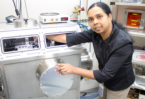
"You can focus the soft X-rays to about 30 nm… more than a thousand times smaller than a human hair," said Chithra Karunakaran, CLS staff scientist for the Soft X-ray Spectromicroscopy (SM) beamline.
Using instruments and a technique (known as STXM) that gives three-dimensional structural information at the nanometer scale, researchers were able to get an understanding of individual quinoa chromosomes, which Karunakaran says "You cannot get using any other instrument."
The level of detail the CLS-Guelph team achieved is ground breaking, she added. The first plant chromosome to ever be studied using a STXM technique was in 1992, but that study was looking at chromosomes more than 10 times the size of quinoa’s. This is the smallest chromosome imaged to date using STXM.
"The results of this study will help to develop new quinoa varieties which may be adaptable to grow in Canadian growth conditions, with disease and virus resistance," said Suresh Neethirajan, University of Guelph bioengineering faculty.
This will be done by developing biomarker libraries, combining information about proteins and genetic material in individual quinoa plants. Using this library, crop researchers can pinpoint where the DNA and protein proportions are unexpected, and from there find the genes tied to everything from cold resistance to certain diseases.
These techniques aren’t limited to helping plants survive, either, “studying chromosomes using nanoscale imaging tools helps to develop novel ways to predict and detect human and crop diseases,” said Neethirajan.
Indeed, the Guelph-CLS team’s technique offers a new opportunity to understand chromosomes in their natural state, without altering them to make imaging easier.
What’s next? Hopefully this technique will enable detailed crop research into the hardiness and diseases of quinoa and other crops, leading to better crops in coming years.
"Even NASA, the US space agency ensures that quinoa is included in the astronaut's diet," said Dr. Neethirajan.
This work was made possible by donations from the Canada Foundation for Innovation, Natural Sciences and Engineering Research Council of Canada, MITACS, University of Guelph, Ontario Ministry of Food and Agriculture, and Canadian Light Source Inc.
Yangquanwei, Z., Neethirajan, S. & Karunakaran, C. Cytogenetic analysis of quinoa chromosomes using nanoscale imaging and spectroscopy techniques. Nanoscale Res Lett 8, 463 (2013). https://doi.org/10.1186/1556-276X-8-463
For more information, contact:
Victoria Schramm
Communications Coordinator
Canadian Light Source
306-657-3516
victoria.schramm@lightsource.ca