
BioXAS-Imaging
BioXAS Imaging beamline is a multi-resolution X-ray fluorescence (XRF) imaging beamline with capabilities to collect XRF imaging data in macro-, micro- and nano-modes. The former two scanning modes are accepting General Users’ proposals and the latter is currently under commissioning.
Spectral Range
Techniques
Imaging & Microscopy
- Macro XAFS/XRF Imaging
- Micro XAFS/XRF Imaging
- Fluorescence Microscopy
Samples
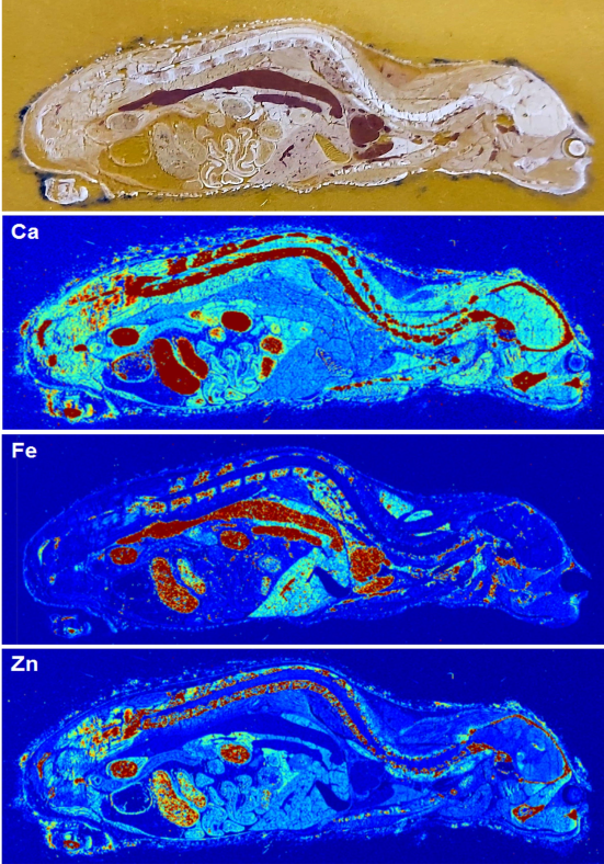
Typical samples used at BioXAS-Imaging are plant or animal tissues, or pieces of rock. On-site capabilities for thin-sectioning samples are available and can be arranged by talking to beamline staff.
Samples are mounted onto holders/frames at a designated area at the BioXAS Imaging beamline.
Specifications
| CLS Port | 07ID-1 |
| Source | In vacuum undulator |
| Typical spot sizes and Photon Flux (@ 100 μm) | Macro-mode 20 µm: 0.6 x 10¹¹ ph/s 50 µm: 3.5 x 10¹¹ ph/s 100 µm: 0.9 x 10¹² ph/s 150 µm: 1.4 x 10¹² Micro-mode 6 x 5 µm (HxV): 1.5 x 10¹¹ ph/s 10 x 5 µm (HxV): 3.2 x 10¹¹ ph/s |
| Resolution | Si(111): 10-4 |
Contacts
Viorica (Ibi) Bondici
Scientist - BioXAS-Imaging
Visit the BioXAS Imaging website for a full list of beamline contacts.
Beamline Video
News Highlights
Loading...
Access
Purchased Access
Purchased access offers quick and accurate solutions to proprietary questions. CLS scientists develop experimental plans based on clients' needs, collect and analyze data, and provide detailed reports with key answers to critical questions.
Peer-Reviewed Access
Academic clients can submit proposals through a peer review process. Beam time is granted based on scientific merit, with the expectation that any results will be published. In special cases, rapid access is also available for instrument or beam time.